
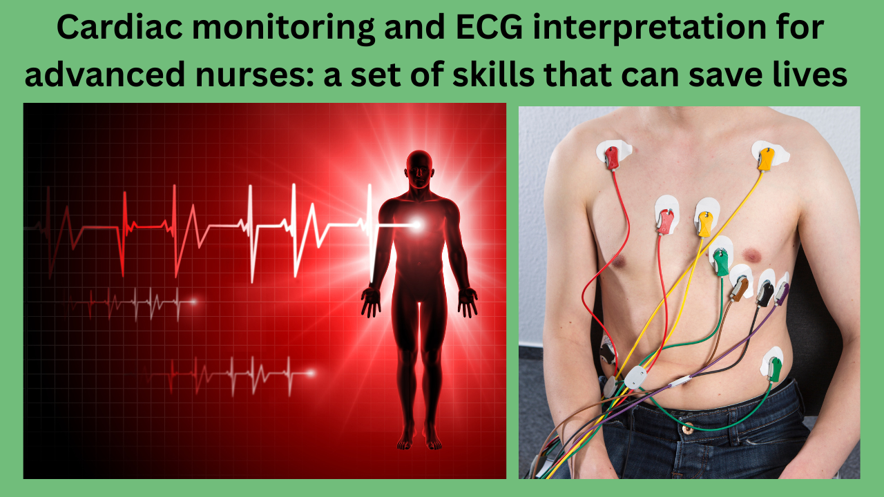
Cardiac monitoring and ECG interpretation for advanced nurses: a set of skills that can save lives
Description: Advanced nurses can learn how to do cardiac monitoring and ECG interpretation. With expert ECG analysis, you can learn to understand heart rhythms, improve emergency care, and raise the level of nursing practice.

All-Inclusive Outline (Table Style)
Heading of Section
Cardiac monitoring and ECG interpretation for advanced nurses: a set of skills that can save lives
Cardiovascular monitoring is an important part of advanced nursing care.
Learning the Basics: What is an ECG, and Why Does It Matter?
Why the heart’s electrical system works the way it does
What an ECG machine is and how it does its job
H2: The Most Important Parts of an ECG Strip
H3 P Wave
H3 PR Time Range
H3 QRS Whole H3 ST Part and T Wave
Do you want a 12-lead ECG or continuous cardiac monitoring?
H3: When to Use Each Kind
H3: Where the Electrodes Are
Heart Rhythms That Every Nurse Should Know
Normal Rhythm of the Sinuses
Atrial Fibrillation (H3)
The third type is ventricular tachycardia and ventricular fibrillation.
H3: Heart Blocks and Brain Fog
H2: A Step-by-Step Guide to Reading an ECG
Rate (H3)
H3 Beat
H3: Axis Difference
Heading 3: Intervals and Shape
ECG Patterns That Put Lives at Risk and How Nurses Respond
H2: Adding Advanced Cardiac Life Support (ACLS)
Monitoring of the heart by nurses in intensive care units
ECG Mistakes That Happen Often and How to Avoid Them
Cardiac monitoring technology uses smart monitors and AI.
Heading 2: Legal and Moral Duty in ECG Monitoring
Questions and Answers (FAQs)
Conclusion 2: The Nurse’s Effect on Heart Survival
You can find out more from the American Heart Association.
H2: Last Word
❤️ Starting off: Cardiac monitoring is an important part of advanced nursing
Heart tracking and reading ECGs are important skills for advanced practice nurses to have in today’s healthcare system. An ECG can quickly identify arrhythmias or cardiac infarctions, potentially saving lives. To be a leader in heart care, nurses need to have the right tools, skills, and ability to think critically.
What is an ECG, and why is it important to know how the heart’s electrical system works?
Electrical signals are what keep the heart beating and contracting. The sinoatrial (SA) node sends these signals, which go through the atria, reach the atrioventricular (AV) nodes, and then go down through the Bundle of His and Purkinje fibers. Arrhythmias can happen if anything goes wrong with this route.
The ECG machine and how it record information
An electrocardiogram, also written as ECG or EKG, is a test that does not hurt the heart and tracks this electrical activity. The ECG leads detect voltage changes and then create tracings on a computer or paper strip to display heart rhythms.
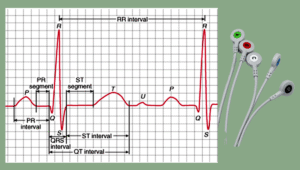
Important Parts of an ECG Strip
For a correct evaluation, it’s important to understand each part of the ECG waveform:
The P wave shows that the atrium is depolarizing. Any issues could indicate the atrium’s growth or the presence of a cardiac rhythm.
PR Time Frame
The PR time frame represents the duration from the onset of the P wave to the onset of the QRS complex. It shows the AV node conduction time.
The QRS complex shows that the ventricle is depolarizing. If the QRS is longer than 0.12 seconds, it means that there are bundle branch blocks or ventricular beats.
Part ST and T Wave
The ST section shows the time between depolarization and repolarization. This kind of elevation or dip often points to myocardial ischemia or infarction. The T wave shows that the ventricles are repolarizing.
Do you want a 12-lead ECG or continuous cardiac monitoring?
How to Use Each One
ECG with 12 leads is a diagnostic tool used for chest pain, fainting, or vital signs that aren’t normal.
Telemetry monitoring is used to monitor a patient’s heart rate when they are critically ill or unstable.
Where the Electrodes Are
The right lead arrangement is critical. Moving things around can lead to false readings and incorrect diagnoses. They need to teach nurses how to put limb and chest leads in the right places.
Heart rhythms that every nurse should know
Normal sinus rate: 60 to 100 beats per minute; steady rate; normal P wave and QRS.
Atrial Fibrillation
The rhythm fluctuates constantly and there are no P waves present. There is a risk of stroke, and the patient requires blood thinners.
Ventricular tachycardia and fibrillation are beats that can kill you. VTach can transform into VFib, necessitating immediate defibrillation.
Heart Blocks and Bradycardia
Problems with the SA node or an AV block frequently lead to slow beats. The patient may require atropine or pacing.
Here is a step-by-step guide on how to read an ECG rate: Please identify the number of QRS complexes in a 6-second strip and then multiply that number by 10.
Rhythm: Check for regularity.
Are P waves present, and is there a QRS following them?
Normal PR Interval: 0.12 to 0.20 seconds.
Normal QRS duration is less than 0.12 seconds.
ST Segment: Search for changes in height or depth.
T Waves: Incorrect patterns can indicate electrolyte imbalance or ischemia.
Life-Threatening ECG Patterns and How the Nurse Responds
In critical care, where there is a lot of stress, nurses must act quickly when an ECG shows changes that could be life-threatening. By noticing these trends, people can act quickly and save lives.
Myocardial Infarction with ST-Elevation
A STEMI occurs when the ST-segment ascends in two or more interconnected leads. Call the cardiac cath lab immediately. Nurses should give air, aspirin, and nitroglycerin, and they should get ready for thrombolysis or percutaneous coronary intervention (PCI).
Point-to-point turns
This type of QT prolongation is often associated with this type of irregular ventricular heartbeat. It includes giving magnesium sulfate intravenously, stopping drugs that extend the QTc interval, and sometimes overdrive pacing.
Asystole, flatline rhythms, and pulseless electrical activity (PEA) require immediate CPR and the identification of reversible causes (H’s and T’s). Epinephrine and effective chest compressions treat PEA and asystole, but they do not treat shockable pulses.
– Adding advanced cardiac life support (ACLS)
When there is a code blue, nurses who are trained in ACLS are critical. The reading of an ECG helps doctors choose which medicines and defibrillation techniques to use.
Tachycardia with a pulse: You might want to think about coordinated cardioversion.
Pulseless rhythms: Do CPR, give adrenaline, and shock.
If you have symptoms of bradycardia, you should start atropine and then transcutaneous pacing if needed.
A favorable ECG reading accelerates the process of making this choice.
Heart Monitoring Led by Nurses in Critical Care Units
Often, critical care nurses are responsible for constant cardiac monitoring. Here are some of their duties:
We are finding the first signs of decompensation.
I am getting in touch with the care team quickly.
We are responsible for educating patients and their families about tracking devices.
The focus is on managing alarm fatigue without jeopardizing patient safety.
They also change the settings on the monitor, move the electrodes, and resolve problems that cause artifacts that look like dangerous beasts.
■ ECG Mistakes That Happen Often and How to Avoid Them
Lead Getting Lost
If you put the leads in the wrong place, especially V1–V6, it can entirely change how you read the ECG. Always stick to the normal landmarks in anatomy.
Interference with electricity (artifact)
Movement, tremors, or loose probes can cause artifact waves, which can resemble VTach or Afib. A beneficial way to secure probes is to teach patients to move as little as possible during readings.
Problems with Machine Calibration
Incorrect gain or speed choices can skew waveforms. When doing 12-lead ECGs, you should always check the accuracy.
Technology used in heart monitoring: smart monitors and AI that work together
These days, watches use AI to analyze the data and automatically find arrhythmias and other problems with the waveforms. Some of the traits are
Telemetry access allows for the remote monitoring of patients at home.
ECG storage in the cloud for analyzing trends.
Predictive algorithms for signs before arrest.
AI aids in decision-making, but nurses are still required to interpret results in light of the patient’s overall health.
📜 Legal and Moral Duties in ECG Monitoring: Advanced nurses need to make sure that
It is crucial to maintain accurate records of ECG events and actions at all times.
Please ensure to obtain informed consent when performing 12-lead ECGs.
It is important to follow HIPAA rules when collecting, keeping, and sharing ECG data.
We are forwarding critical data immediately to the physician team.
Not noticing and reporting changes can lead to lawsuits and bad results for patients.
Frequently Asked Questions about Heart Monitoring and Reading ECGs
1. What is the normal range for a PR interval?
The average time between PRs is between 0.12 and 0.20 seconds. Longer gaps could be a sign of a first-degree atrioventricular (AV) block.
2. How often should you switch out the sensor leads?
Every 24 to 48 hours, telemetry lines should be moved and cleaned to keep the signal clear and stop the skin from breaking down.
3. Can an ECG help a nurse tell if a patient has an arrhythmia?
Even though nurses can’t make official diagnoses, they can spot problems, write them down, and ask the provider to make a diagnosis.
4. What does a T wave that has a peak mean?
A tall, peaking T wave could mean that you have hyperkalemia, an electrolyte excess that can be life-threatening.
5. What can nurses do to cut down on false warnings in heart monitoring?
Make sure the lead is in the right place, keep the skin clean, and change the sensitivity settings based on how the patient moves and their baseline beat.
6. What’s the difference between an ECG and telemetry?
Telemetry allows you to continuously monitor your heartbeat. An ECG with 12 leads gives a more complete picture of the heart’s electrical activity.
Conclusion: The Nurse’s Effect on Heart Survival
Interpreting an ECG and monitoring the heart are not merely technical skills; they have the potential to save lives. Nurses are often the first to notice small but important changes in a patient’s heart rhythm because they pay close attention, understand, and use their clinical expertise.
Just desire to say your article is as astonishing.
The clarity for your publish is simply excellent and that i could think
you are knowledgeable in this subject. Well along with your permission let me to take hold of your RSS feed to stay updated with forthcoming post.
Thank you one million and please continue the rewarding work.
Thank a lot for your Appreciation and Motivation 🙏🙏🙏🙏🙏